Course Content
There are 7 lessons in this course:
- Cytology
- Common Organelles & Structures such as Plasma Membrane, Cytoplasm, Cytosol, Cytoskeleton, Nucleus, Endoplasmic Reticulum (ER), Mitochondria, Golgi Apparatus, Ribosomes, Lysosome and Peroxisome
- Specialised Organelles & Structures - Sarcolemma, Sarcoplasmic Reticulum, T-Tubules, Undulipodia and Microvilli
- The Anatomy of Cellular Division - Review, Characteristic Interphase Structures, Characteristic Mitosis Structures.
- Surface Anatomy
- Anatomy
- Terminology: Regions and Positions of the Body, Planes and Views of the Body, Locational and Directional Terminology, Descriptive Terms, Numerical Terms
- Surface Anatomy
- Superficial Structures, Features and Markings - the Head, the Neck, the Back, the Anterior Torso, the Upper Extremities and the Lower Extremities.
- Practical Use of Surface Anatomy - Inspection, Palpation, Auscultation, Percussion, Measuring Vital Signs, Blood Testing and Signs versus Symptoms.
- Systemic Anatomy I
- Systemic Anatomy - Integumentary, Nervous, Endocrine, Immune etc.
- Organisation of the Body
- The integumentary System - Anatomy of the Skin and Anatomy of other Integumentary System Components
- The Nervous System - the Brain, the Spinal Cord, Spinal Nerve Anatomy, Divisions of the Nervous System
- The Cardiovascular System - the Heart and the Vasculature
- The Renal System - the Kidneys, the Ureters, the Bladder and the Urethra.
- Systemic Anatomy II
- The Endocrine System - the Pineal Gland, the Pituitary Gland, the Hypothalamus, Thyroid, Parathyroids, Thymus, Pancreas, Adrenal Glands and Ovaries and Testes.
- The Immune System - Thymus, Spleen, Bone Marrow and Lymphatic System
- The Male Reproductive System - External Components and Spermatogenesis in the Seminiferous Tubules of the Testes.
- The Female Reproductive System - the Uterus, the Vagina, the External Genitalia, the Ovaries and Fallopian Tubes, Accessory Glands and the Breast and Mammary Tissue.
- Regional Anatomy I
- Regional Anatomy - Integument, Peripheral Nervous System, Vasculature, Skeleton, Musculature
- Musculature - Brevi, Extensor, Indicis, Longus, Palmar etc.
- Bone - Girdle, Notch, Spine, Tuberosity etc.
- Vasculature and Miscellaneous - Axilla, Cubital, Palpate etc.
- The Cranial Cavity - Bones of the Skull and Facial Bones
- Thoracic Cavity - the Oral and Nasal Cavities, the Paranasal Sinuses, the Nasopharynx, the Oropharynx, the Laryngopharynx, the Larynx, Laryngeal Membranes, Ligaments and Muscles, the Trachea, the Bronchial Tree, the Lungs and the Diaphragm
- The Abdominopelvic Cavity - the Oesophagus, the Stomach, the Small Intestine and the Large Intestine.
- Regional Anatomy II
- The Upper Extremities - Anatomical features of - the Humerus, the Ulna, the Radius, the Carpals, the Metacarpals, the Phalanges; Musculature of the Upper Extremities, Innervation of the Upper Extremity, Vasculature of the Upper Extremity.
- The Lower Extremities - Anatomical Features of - the Pelvis, the Femur, the Tibia, the Fibula, the Tarsals, the Metatarsals, the Phalanges, Innervation of the Upper Extremity and Vasculature of the Lower Extremities.
- Radiographic and Diagnostic Anatomy
- Medical Imaging - X-Ray, CAT scan, MRI, PET Scan and Ultrasound
- Diagnostic Anatomy - Abdominopelvic Cavity, Abdominopelvic Quadrants.
Each lesson culminates in an assignment which is submitted to the school, marked by the school's tutors and returned to you with any relevant suggestions, comments, and if necessary, extra reading.
Aims
- Describe and understand the microscopic anatomical features of human cells.
- Review basic structure and form markings of the body and be able to name them.
- Describe the significant systems and the structure of those systems of the body: Integumentary, Nervous, Cardiovascular and Renal Systems.
- Describe the significant systems and the structure of those systems of the body: Endocrine, Immune and Reproductive Systems.
- Describe the significant structures in specific compartments or parts of the body: body cavities.
- Investigate the anatomy of the extremities - the arms and legs. This includes the bone, musculature and nervous tissue of the regions.
- Describe the study of the structure of the body and the application of various forms of medical imaging.
What will you do in the course?
- Describe the importance of the following structures of the eye: eyelids, eyelashes, and eyebrows.
- What structures form the oral cavity? Briefly describe their importance.
- Using the internet or other reference material, outline and describe otitis media and its causes.
- Besides the eyes, ear, and mouth - what other structures can be studied without a microscope ? List at least ten.
- Using the internet or other reference material, describe the three basic functions of the nervous system that are necessary to maintain homeostasis.
- Using reference materials or the internet, distinguish between grey and white matter and describe where they are found and their differences.
- Using the internet or other reference material define the following: resting membrane potential, depolarization, repolarization, polarized membrane, nerve impulse, depolarized membrane, repolarized membrane, and refractory period.
- List and describe the structure of the four principle parts of the brain.
- Compare and contrast neurons and neuroglia, describing both structure and function.
- List the names and locations of the principal body cavities and their major organs.
- List the names and locations of the abdominopelvic quadrants and regions.
- In which quadrant would you feel the pain from appendicitis? From an inflamed liver or gallbladder problems? Problems with the sigmoid colon? Problems with the spleen?
- Using the internet or other reference materials find a sample image of the listed medical imaging techniques.
This course assumes a general background knowledge of Anatomy and Physiology, equivalent to Human Biology IA or similar. An additional textbook is not required, however if you wish to purchase one please contact the school for advice. This course is specifically concerned with Anatomy and there is very limited discussion of physiology. Our companion course, Physiology II may be taken following this module to enhance your understanding of this subject area.
In this course:
Before we investigate the gross anatomical structures of the body, it is important to understand the individual units that make up all tissues; cells. Many vital processes occur in the cell. Different cells have different structures, numbers of organelles and so on, to enable them to perform their specific functions. These cells together form tissues which in turn group to form organs and organ systems that combine different cell types to enable systems to perform coordinated functions.
In order to investigate systemic and regional anatomy later in the course, it is first important that we look at the microscopic anatomy of cells. We will start by discussing common organelles and structures, before examining some of the specialised structures found in some cell types.
The study of the human body can be divided into specific fields, one of which is anatomy. Anatomy is the study of structure, how parts of the body are sized and shaped and how they interact with each other, as well as the tissues that form them. It does not consider how parts of the body function; what they do, this is the field of physiology.
Anatomy is and was the starting point of scientific investigation of the human body. Without an understanding of structure we cannot fully understand function, for it is the structure and interrelation of body parts that permits their function. In order to study anatomy, it is important to understand the different medical/scientific terms that are used to indicate location, relationship, components, numbers and so on. Key terms are listed in the following tables, some will be familiar, but should be reviewed along with new terms, to ensure you are able to fully understand this course.
DO YOU UNDERSTAND SURFACE ANATOMY?
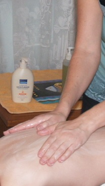 Surface anatomy is, as the name suggests, the study of the anatomical features, tissues, organs and structures found on the surface of the body. It also includes those near to the surface, that can be examined from the surface either visually, audibly or by palpation. In order to make investigations of structures knowledge of the surface projection of the structure, or its location relevant to surface features is essential. For example, the positioning of a stethoscope on the chest wall so as to maximise the audition of heart sounds.
SUPERFICIAL STRUCTURES, FEATURES AND MARKINGS
The Head
Nose
The nose forms a ridge that sits proud of the face. Medial to the eyes is the bridge of the nose, and it feels hard because it lies over the nasal bones. The softer fleshy inferior part of the nose ridge is the dorsum nasi. The most inferior medial tip of the nose is the apex. The two orifices at the inferior of the nose are the nostrils (or external nares), which are separated medially by the nasal septum, and bordered laterally by the ala nasi.
Mouth
The mouth is inferior to the nose, and is composed of the upper and lower lips (labia) which are a darker pink colour compared to the surrounding skin. The grooves running superiorly from each side of the mouth to the lateral side of the nostrils are the nasolabial sulci
Philtrum
This is the region intermediate to the nose and upper lip. It is characterised by two parallel vertical soft ridges running to the lip from each nostril with an intermediate depression.
Eyes
The orbital area includes both eyes, with their protective superficial eyelids (superior palpebra) and the inferior palpebra. The superior palpebral fissure is the crease formed by the upper eyelid (absent in people of Asian background). Eyelashes are found on the palpebra. Superior to the eyes is the superciliary arch, which is defined by horizontal, curved eyebrows.
Frontal region
This is the relatively featureless area immediately superior to the superciliary arches, commonly known as the forehead.
Buccal region
These are the rounded lateral features of the face, commonly known as the cheeks. The mouth and the cheeks form the oral region of the face.
Mental region
This is the region inferior to the mouth that is commonly known as the jaw, or chin (mentum). The mentum tends to have a sharper angle in females, and to be squarer in males.
Ears
The auricular area of the face. The ears are located laterally and have a superior cartilaginous region, and a soft fleshy pinna (or auricle). The orifice in each ear is the external auditory canal. Posterior to the ears and slightly inferior is a palpable bony bump known as the mastoid process.
Posterior skull features
On the inferior posterior of the head is a palpable bony ridge known as the external occipital protuberance.
The eyes, ears, nose and mouth will be discussed in detail in Lesson 5 (Regional Anatomy I)
The Neck
Many of the structures of the neck can be palpated, including:
The trachea
Felt along the midline as a series of horizontal bumps
The larynx
Felt likewise in the superior portion of the neck
The Adam’s apple
A more prominent visible bump in males and is formed by the thyroid cartilages
Vertebra prominens
This is the palpable bony bump felt medially at the posterior of the inferior base of the neck.
Sternocleidomastoid muscles
This runs vertically and slightly obliquely down the neck. They form two surface triangles, the anterior and posterior triangles, which are further subdivided into smaller triangles.
Anterior Triangle
- The submental triangle is the most superior and sits under the chin. A doctor will palpate here to feel for swollen lymph nodes, indicative of infection.
- The submandibular triangle sits lateral to the submental triangle.
- The carotid triangle is inferior to the submandibular triangle. Pulse rate can be measured by palpating in this region, as the carotid arteries run through it.
- The muscular triangle is inferior to the carotid triangle
Posterior Triangle
- The occipital triangle forms the superior portion of the posterior triangle.
- The supraclavicular triangle is inferior to the occipital triangle, finishing along the superior border or the clavicle. Some portions of the large veins and arteries serving the upper extremities run through this region.
The Back
Medial furrow
This is a valley in the back that as the name suggests runs along the midline of the torso. It can be divided into the cervical furrow, the thoracic furrow, which is broad and shallow and the lumbar furrow which is a deeper furrow. The deep groove between the buttocks is the lumbar furrow, deep to which is the coccyx. Palpating along the furrow you can feel the spinous processes of the vertebrae making up the vertebral column. When a person flexes at the hip (bends over forward) the superior portions of the furrow flatten, and the spinous processes in the region become clearly visible as bumps along the midline.
Inferior angle of Scapula
In the superior portion of the back, lateral to the medial furrow are two protruding points, one on either side that are often visible and can be easily felt. These are formed by the inferior portion of the scapula (shoulder blade).
Spine of Scapula
Superior to the inferior angle of the scapula, approximately level with the shoulders are slightly protruding lines running horizontally. These are formed by the spine of the scapula (shoulder blade).
Gluteal Muscles
The gluteal muscles form the shape of the buttocks. The most superficial of them, the gluteus maximus gives the bulk to the buttocks.
The Anterior Torso
Ribcage and Intercostal Spaces
There are 12 sets of ribs in the average person’s chest. The superior 7 pairs are the true ribs that have their own cartilaginous joint; the intermediate 3 pairs are the false ribs, sharing a single cartilaginous joint to the sternum. The most inferior 2 pairs are the floating ribs, that attach to the vertebrae only, and not the sternum. Superficially, the ribs form rounded ridges that run laterally from the midline. Between the ribs are shallow (or in malnourished people quite deep) depressions which are superficial to the intercostal spaces.
Pectoralis Major and Nipples
The two pectoralis muscles sit contra-laterally in the upper torso. Superficially they protrude slightly to give the ‘pecs’. In women, they are deep to the mammary tissue and are not prominent. They are more prominent in muscular individuals ad in obese men may be obscured by more superficial fat deposits. Superficially, on the inferior of the raised area of the pecs or the breasts are two contralateral darkened circular regions of tissue. They are comprised of an outer-most flat area known as the areolar and a proud central area known as the nipple.
Sternum (body)
The body of the sternum runs along the midline, slightly depressed compared to the lateral pectoralis major muscles, or in women, the breasts.
Clavicles and Associated Fossas
The two clavicles form rounded ridges that sit proud of the surrounding structures, running laterally towards the shoulders. Superior to each clavicle is a small depression known as the supraclavicular fossa. Inferior to the medial portion of each clavicle is another small depression known as the infraclavicular fossa. Medial to the two heads of the clavicles is a deeper depression bordered by the sternocleidomastoid muscles, this is the suprasternal notch.
Costal angle
The angle formed immediately inferior to the xyphoid process by the most distal pair of ribs. An upside down V shape that is readily visible on a very thin person.
Rectus abdominis
This is the major superficial muscle of the abdomen. It is a paired muscle, running down the midline and separated by the linea alba. It runs from the xyphoid process to the pubic bone of the pelvis and bulges slightly in muscular individuals to give the ‘6 pack’.
Umbilicus
This is the depression (or in some cases projection) in middle of the abdomen where the umbilical cord once entered the foetal body in utero. It is commonly known as the belly button and is used to mark out abdominopelvic quadrants.
Anterior superior iliac spine
The spine is an anatomical feature of the lateral bones of the pelvis (the iliac bones). Superficially they can be palpated and in slimmer individuals may obviously protrude.
Iliac crest
The iliac crest runs intermediate to the anterior and posterior iliac spines of the pelvis. It can be palpated and followed from the anterior superior iliac crest. This is commonly known as the hip.
The Upper Extremities
Deltoid Muscle
The proximal rounded shape of the upper arm is produced by the underlying deltoid muscle.
Axilla
The region under the upper arm commonly known as the armpit. May be covered by hair in adults. Characterised by two folds, the anterior axillary fold, formed by the pectoralis major, and the posterior axillary fold formed by the teres major and latissimus dorsi muscles.
Biceps brachii
This is the major anterior muscle of the upper arm. It can be palpated and forms a rounded bulge in muscular individuals.
Antecubital or Cubital Fossa
The anterior surface over the elbow joint. The brachial vein runs superficially over the joint in the antecubital fossa, and is punctured to draw blood. It is often visible as a blue line.
The Elbow
The prominence that forms the (posterior) elbow is the olecranon of the ulna. IT can be palpated when the arm is either straight or bent. When the arm is straight the skin overlying the joint is wrinkled.
Anatomical snuff box
A depression proximal to the thumb, most obvious when the thumb is maximally abducted. Within this region the pulse may be taken using the radial artery and the scaphoid bone palpated.
The Wrist
The small medial prominence on the anterior of the wrist is made by the underlying pisiform bone of the wrist. Posteriorly, on the medial surface is a second small rounded prominence made by the underlying styloid process of the ulna. Anteriorly, a small ridge running medially to the palm is the tendon of the palmaris longus muscle. Lateral to that is a smaller ridge formed by the underlying tendon of the flexor carpi radialis.
The Knuckles
The knuckles are immediately proximal to the fingers and are rounded prominences formed by the heads of the metacarpal bones.
Thenar and Hypothenar Eminences
The bulky rounded region medial to the thumb on the palm is the thenar eminence; the smaller rounded prominence proximal to the little finger is the hypothenar eminence. They are both muscular formations.
WHAT SETS ACS APART?
At ACS we provide you with more than just a set of course notes.
Your 'learning package' includes:
- Course notes.
- Self-assessment quizzes.
- Assignment feedback.
- You can interact one on one with a professional tutor with decades of experience - just email, phone or log on to chat to connect with them.
- Depending upon your course, your studies may involve independent research, interviews, practical exercises, assessments, Problem Based Learning projects, and more.
WHAT NEXT?
Register to Study - Go to “It’s Easy to Enrol” box at the top of the page and you can enrol now.
or
Get Advice – Email us at info@acsedu.co.uk OR
Use our FREE COUNSELLING SERVICE to contact a tutor
CLICK TO CONTACT US