Types of Microscopes
 Microscopes are essential for study in biological science. There are many different types of microscopes; and each has it's uses.
The cheapest and most common microscopes are "light microcopes"; but these are limited as to how much detail they can see. Other microscopes can look at far more detail; but they are extremely expensive to build or buy.
Light Microscopes
There are three types of light microscopes that allow you to see small things by bouncing light off those subjects.
- Stereoscope (or Stereo Microscope)
- Compound Microscope
- Confocal Microscope
Stereoscope (or Stereo Microscope)
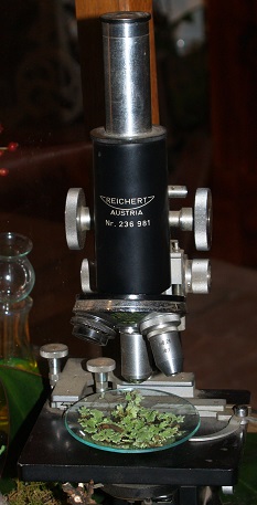
A stereo microscope is widely used in science. It allows for a magnified image to be viewed and is predominantly used for studying larger specimens as there is a limitation to the strength of magnification. For example, it is ideal for studying insects, plant material, some fungal samples and soil samples. Its use in microbiological applications is extremely limited as it doesn’t off resolution or magnification to adequately view micro-organisms.
Compound Microscope
A compound microscope includes a number of lenses and is a commonly used microbiology laboratories. The light enters from the base of the microscope through the condenser, through the specimen on a glass slide into the objective lens. The objective and ocular lens magnify the image before it can be seen through the eye piece. The light passing through the specimen can be controlled by the iris diaphragm. When using objective lens of higher magnification, more light is required to see the image clearly.
Dark-Field Microscopy is used to examine organisms that are light-sensitive and are better seen with a darker background. This microscope contains a condenser that prevents light being transmitted directly through the specimen, but causes the light to be reflected at an angle. The image seen is of a light specimen on a dark background.
Phase-Contrast Microscopy is generally required to observe micro-organisms live and unstained. Stains that are generally used to observe micro-organisms cause cell-death, and therefore they cannot be used on live samples. A phase-contrast microscope is fitted with a specialised condenser and objective lens so that the slightest changes in refracted light can be used to observe live specimens.
Fluorescence Microscopy is routinely used in diagnostic laboratories for identification purposes, whereby the fluorescent molecules are excited using ultraviolet light. Fluorescent dye molecules are attached (or tagged) to Antibody molecules, and will combine with Antigens if present in a specimen. Multiplexing of a number of fluorescence dyes can be used in these methods.
Confocal Microscope
Confocal microscopy is carried out through the use of ultraviolet laser beams that can be targeted through very small holes or slits. The fluorescence is then tightly focused, with the imagine being reconstructed by a computer to provide a high resolution image. A confocal microscope can pass through a thicker specimen at successive planes, providing three-dimensional images.
ELECTRON MICROSCOPY
Electron microscopy has moved from using light beams through glass lenses, to the combining the use of beams of electrons and electromagnets to visualise very small structures. It is necessary to have the electrons moving through a vacuum, this removal of air allows for high resolution images to be produced at extremely high magnifications.
There are two common types of electron microscope:
- Scanning Electron Microscope (SEM)
- Transmission Electron Microscope (TEM)
Scanning Electron Microscope (SEM)
SEM is used to image the surface of micro-organisms. The specimen to be examined is first coated with gold; then electrons are fired onto the surface and bounced off to a sensor that records a three-dimensional image.
Transmission Electron Microscope (TEM)
TEM is used predominantly to visualise the internal structures of micro-organisms. Specimens to be used with TEM need to be embedded into plastic and cut into thin sections (70-90nm). These are then transferred onto a thin wire grid and placed in front of the electron beam.
Particularly small specimens (for example viruses) can undergo a technique called Shadow casting, whereby the thin sections are coated with a heavy metal such as gold. This technique provides a three-dimensional effect to the image, as a “shadow” will be seen where there is no heavy metal present (that is behind the specimen, which was an area blocked from coating).
HELIUM ION MICROSCOPE
More recently advancements have been made in microscopy with the development of the Helium Ion Microscope (HIM). The helium gas is used at very low temperatures, where it intensifies the electrical field at the tip of a sharpened needle. This process allows for a very bright ion beam to be formed. After passing through three different detectors, an image is acquired that provides more information compared to SEM. It enables material to be seen at atomic level. HIM is not limited by the gold coating process of SEM and provides quality images with high resolution, sensitivity and increased structural detail.
[17/07/2026 13:15:41]